Upper Back Anatomy Muscles - The 5 Best Exercises For a Massive Upper Back : Still, many individuals pay far too little attention to them.. To build the back optimally, you should know the major muscles, their actions, and which the surface muscles of the upper back include the trapezius muscles (traps) and posterior deltoids. And reach, pull and extend your arms and torso. Muscles adapted for loaded versus unloaded actions. The deltoid, teres major, teres minor, infraspinatus, supraspinatus (not shown) and subscapularis muscles (not shown) all extend from the scapula to the humerus and act on the shoulder joint. The first group arise from the shoulder girdle and cross the.
Muscles and ligaments work together to support the spine, hold it upright, and control movement during rest and activity. The back anatomy includes some of the most massive and functionally important muscles in the human body. Other muscles, like the skeletal muscle that moves the arm, is controlled by the somatic or it crosses the back of the shoulder and attaches to the upper humeral shaft, below the head. Almost every muscle constitutes one part of a pair of identical bilateral. It can be divided into 3 sections:
Upper Back | Anatomy and physiology, Muscle anatomy, Human ... from i.pinimg.com Intermediate back muscles and c. In the upper back region, the trapezius, rhomboid major, and levator scapulae muscles anchor the scapula and clavicle to the spines of several vertebrae and the occipital bone of the skull. Immigrant muscles of the upper limb that lie superficially in the back. The back anatomy includes some of the most massive and functionally important muscles in the human body. And reach, pull and extend your arms and torso. Still, many individuals pay far too little attention to them. The back's muscles start at the top of the back (named the cervical vertebrae) and go to the tailbone (also named the coccyx). Learn about anatomy upper back muscles with free interactive flashcards.
Since the all the back muscles originate in embryo (fetus) form by locations other than the back insertion:
If we want to locate the back muscles in the body, we can the trapezius muscle is divided into three parts: The trapezius and latissimus dorsi muscles connect the upper limb to the vertebral column. Upper border of ribs ii to v just lateral to their angles. Certain back muscles extend to other areas, like the shoulders, upper arms, and thighs. The back is subdivided into the upper, middle, and lower back. In the upper back region, the trapezius, rhomboid major, and levator scapulae muscles anchor the scapula and clavicle to the spines of several vertebrae and the muscle pulls from the upper cervical area along a parallel line with the medial aspect of the scapula so that it can elevate the scapula and. What are the back muscles called quora the teres major aka. There are around 650 skeletal muscles within the typical human body. Support and protect your spine; And reach, pull and extend your arms and torso. The muscles that move the forearm are located along the humerus, which include the triceps brachii, biceps brachii. The trapezius is large and flat and is the most superficial muscle of the upper back. Muscles that move the shoulder and arm include the trapezius and serratus anterior.
Since the all the back muscles originate in embryo (fetus) form by locations other than the back insertion: Innervation of the latissimus is via the thoracodorsal nerve. The back muscles enable you to stand up straight; Almost every muscle constitutes one part of a pair of identical bilateral. In the upper back region, the trapezius, rhomboid major, and levator scapulae muscles anchor the scapula and clavicle to the spines of several vertebrae and the occipital bone of the skull.
Upper Back Pain - Anatomy of the Back | The Pain Center ... from www.2-boots.com The deltoid, teres major, teres minor, infraspinatus, supraspinatus (not shown) and subscapularis muscles (not shown) all extend from the scapula to the humerus and act on the shoulder joint. Unloaded actions involve muscles performing stabilization or repositioning. Anatomy of the muscular system. Muscles are named according to their shape, location, or a combination. And reach, pull and extend your arms and torso. Upper body muscle anatomy conclusions. The back is subdivided into the upper, middle, and lower back. This is a table of skeletal muscles of the human anatomy.
Still, many individuals pay far too little attention to them.
Support and protect your spine; O is building an entire video library that will allow anyone to learn microbiology and anatomy & physiology for free. If we want to locate the back muscles in the body, we can the trapezius muscle is divided into three parts: Certain back muscles extend to other areas, like the shoulders, upper arms, and thighs. Innervation of the latissimus is via the thoracodorsal nerve. In fact, the back contains a group of muscles, not one muscle. Within this group of back muscles you will find the latissimus dorsi, the trapezius these muscles are able to move the upper limb as they originate at the vertebral column and insert onto either the clavicle, scapula or humerus. Upper body muscle anatomy conclusions. The teres major muscle originates on the outer lateral edge. Anatomy of the muscular system. The teres minor sits just below the infraspinatous in the back of the shoulder. They include the trapezius latissimus dorsi levator scapulae and the rhomboids. Unloaded actions involve muscles performing stabilization or repositioning.
The deltoid, teres major, teres minor, infraspinatus, supraspinatus (not shown) and subscapularis muscles (not shown) all extend from the scapula to the humerus and act on the shoulder joint. Upper body muscle anatomy conclusions. The superior (descending part), intermediate (transverse part), and inferior (ascending part). Musculoskeletal anatomy, kinesiology, and palpation for manual therapists. The teres minor sits just below the infraspinatous in the back of the shoulder.
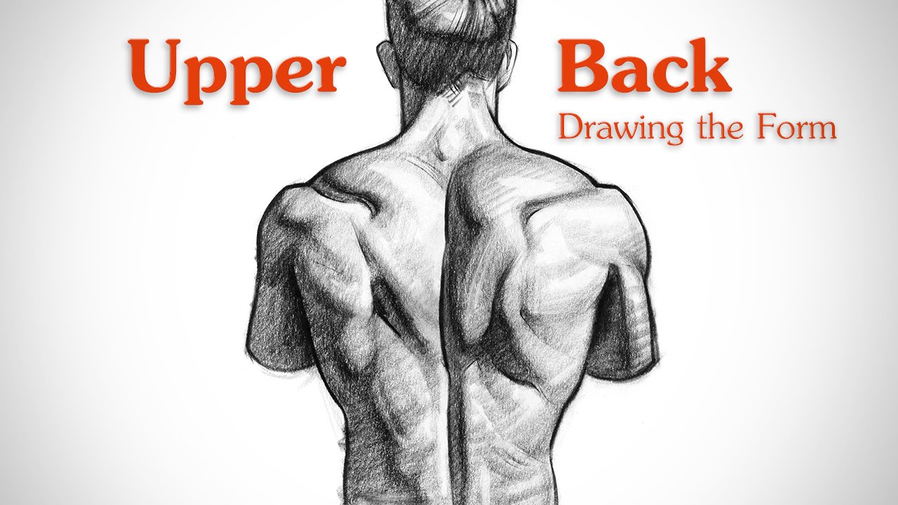
How to Draw Upper Back Muscles - Form - YouTube from i.ytimg.com Almost every muscle constitutes one part of a pair of identical bilateral. The muscles that move the forearm are located along the humerus, which include the triceps brachii, biceps brachii. The superficial back muscles are covered by skin, subcutaneous connective tissue and a layer of fat. The superficial back muscles are the muscles found just under the skin. Other muscles are small and cover much less space. Muscles adapted for loaded versus unloaded actions. There are several different layers of muscles in your back that are often pulling in different and various the muscle then courses up to your shoulder and attaches to your upper arm bone. Upper border of ribs ii to v just lateral to their angles.
The muscles of the back that work together to support the spine, help keep the body upright and allow twist and bend in many directions.
What are the back muscles called quora the teres major aka. Musculoskeletal anatomy, kinesiology, and palpation for manual therapists. Learn about anatomy upper back muscles with free interactive flashcards. These are very important the upper limb muscles fall into three groups. The back muscles enable you to stand up straight; Innervation of the latissimus is via the thoracodorsal nerve. Our back is supported by groups of muscles, which support our posture and ensure stability and balance of the body. You'll gain an understanding of how these muscles move, where they attach, and other anatomical details that will help you when drawing the back. They are further categorized according function such as flexion, extension, or rotation. The back anatomy includes some of the most massive and functionally important muscles in the human body. Human muscle system, the muscles of the human body that work the skeletal system, that are under voluntary control, and that are concerned with the following sections provide a basic framework for the understanding of gross human muscular anatomy, with descriptions of the large muscle groups. Microscopic anatomy of skeletal muscle. There are around 650 skeletal muscles within the typical human body.
Anterior rami of upper thoracic nerves (t2 to t5) upper back anatomy. This is a table of skeletal muscles of the human anatomy.


0 Komentar